Health warning: The most potent "liver-damaging" drink, causing cancer, loved by both adults and children
Tips 22/12/2025 16:32
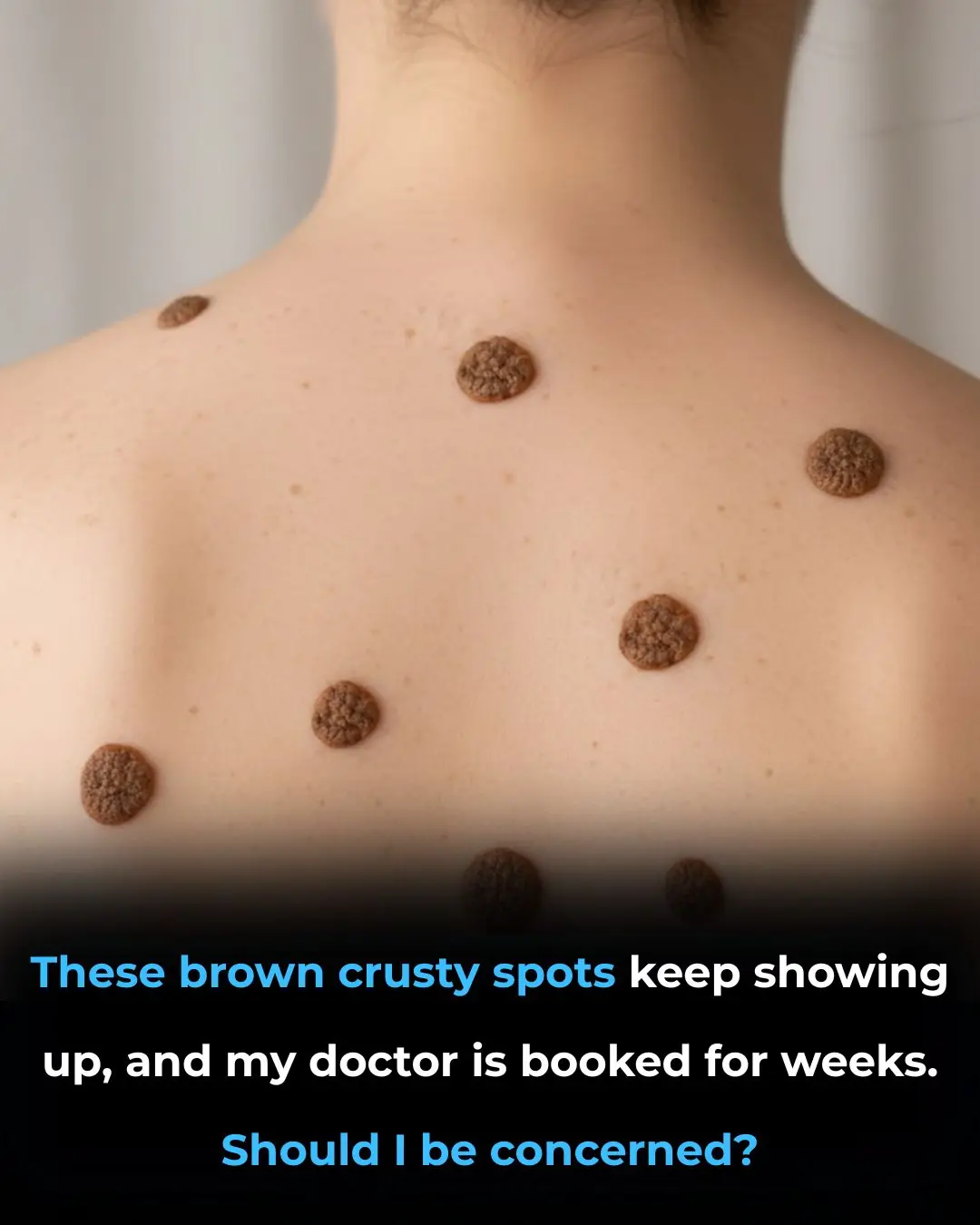
Noticing sudden changes on your skin can be unsettling, especially when brown, crusty spots seem to appear without warning. These changes often trigger concerns about skin cancer, premature aging, or underlying health problems. The situation can feel even more stressful when dermatology appointments are difficult to secure and long waiting times leave many people searching for answers on their own.
This article is designed to help ease that uncertainty. We will explain what these brown, crusty spots are most commonly associated with, how to recognize signs that may be harmless versus those that deserve medical attention, and what you can safely do while waiting to see a dermatologist. By understanding these skin changes, you can approach your skin health with greater confidence and responsibility.
In many cases, brown, crusty spots on the skin are seborrheic keratoses. These are extremely common, non-cancerous skin growths that usually develop in adults over the age of 40, though they can appear earlier. Their color ranges from light tan to deep brown or black, and their surface often looks dry, rough, or wart-like.
Seborrheic keratoses can be tiny or grow larger over time, sometimes exceeding an inch in diameter. While they may look alarming, they are considered benign and usually do not pose a health risk. Treatment is generally optional unless the growth becomes irritated, catches on clothing, or causes cosmetic concern.
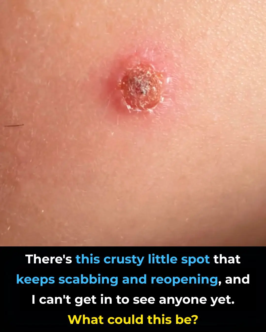
Telling the difference between seborrheic keratoses and skin cancer can be difficult without professional training. Seborrheic keratoses typically have a waxy or scaly texture and appear as though they are sitting on top of the skin. In contrast, melanoma often has uneven borders, multiple colors, and an asymmetrical shape.
Other skin cancers, such as basal cell carcinoma or squamous cell carcinoma, may appear as sores that do not heal, red or scaly patches, or firm bumps. Any lesion that bleeds easily, becomes painful, or changes rapidly should be evaluated by a medical professional as soon as possible.
One of the most distinctive features of seborrheic keratoses is their “stuck-on” look. They often resemble drops of wax or dried clay that seem pasted onto the skin rather than growing from within it. This characteristic alone helps dermatologists identify them quickly.
The surface texture can range from smooth and slightly raised to thick, crusty, or wart-like. Colors may vary widely, even within the same person. While this variety is normal, any dramatic or sudden change should still be monitored carefully.
Seborrheic keratoses become more common with age and are especially prevalent in middle-aged and older adults. Genetics play a major role—if close family members have them, you are more likely to develop them as well. Sun exposure, skin friction, and hormonal factors may also contribute.
It is normal for these spots to increase in number over the years. They may appear gradually or in clusters, sometimes giving the impression that they developed overnight. In most cases, this increase is part of the natural aging process rather than a sign of disease.
Although most brown skin growths are harmless, certain warning signs should never be ignored. A spot that changes shape, develops irregular borders, shows multiple colors, or grows rapidly requires medical attention. Bleeding, pain, or persistent itching are also red flags.
Another concerning sign is the sudden appearance of many new lesions in a short period of time. While this is often benign, it can occasionally signal an internal medical issue. When in doubt, it is always safer to consult a healthcare provider.
For stable, symptom-free seborrheic keratoses, waiting for a scheduled dermatology visit is usually safe. These lesions do not typically require emergency care. However, if you notice alarming symptoms such as bleeding, ulceration, rapid growth, or significant discomfort, you should seek medical evaluation sooner.
Individuals with a personal or family history of skin cancer should be especially cautious and proactive. Early evaluation can significantly improve outcomes if a serious condition is present.
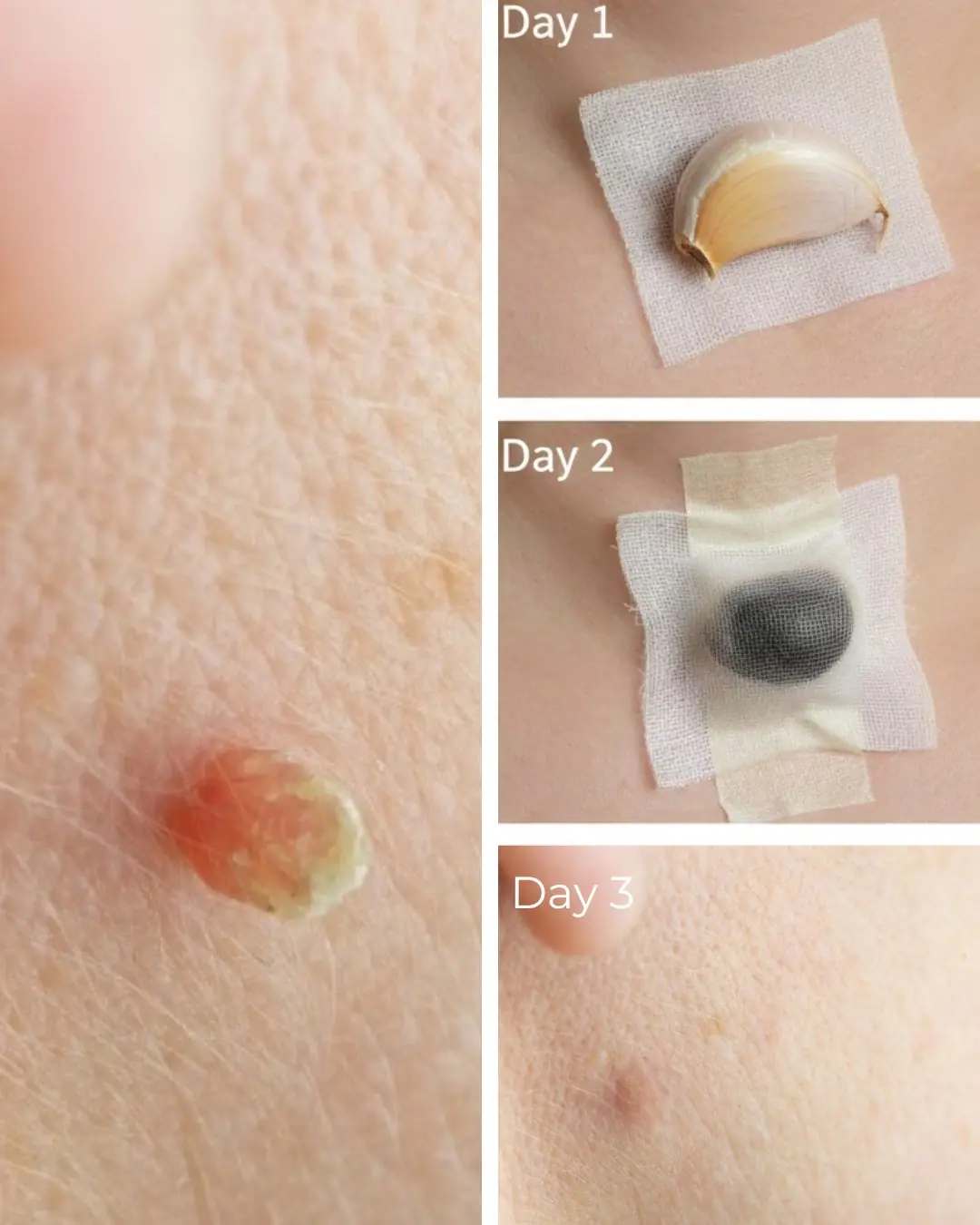
Social media platforms are full of home remedies and “quick fixes” for removing skin growths, but these methods can be dangerous. Attempting to burn, cut, or chemically remove skin lesions at home can lead to infection, scarring, or delayed diagnosis of skin cancer.
Without proper examination, it is impossible to know whether a lesion is truly harmless. Dermatologists use sterile tools and controlled techniques to ensure both safety and accurate diagnosis—something DIY methods cannot provide.
During a dermatology visit, the doctor will perform a full skin examination and may use a dermatoscope to closely inspect the lesion. This handheld device allows the dermatologist to see patterns and structures beneath the skin’s surface.
If there is any uncertainty, a biopsy may be performed. This involves removing a small sample of the lesion for laboratory testing. Biopsies are usually quick, minimally uncomfortable, and provide definitive answers.
Seborrheic keratoses do not need to be removed unless they cause discomfort or cosmetic concern. When removal is desired, several safe options are available. Cryotherapy uses liquid nitrogen to freeze the growth, while curettage involves gently scraping it off.
Electrosurgery may also be used to destroy the lesion with electrical energy. Your dermatologist will recommend the most appropriate method based on the size, location, and characteristics of the spot.
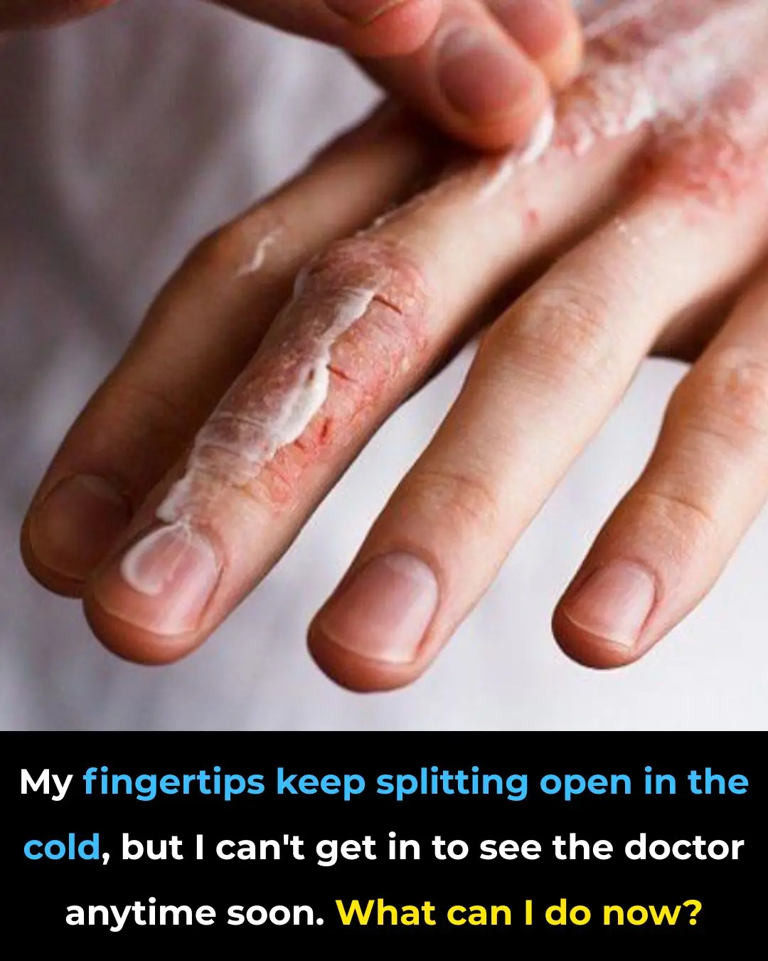
While waiting for your appointment, performing regular self-examinations can be very helpful. Use mirrors or ask for assistance to check areas that are difficult to see. Pay attention to any changes in size, color, texture, or sensation.
Keeping notes or taking photos over time can help track changes accurately. Additionally, protecting your skin with broad-spectrum sunscreen (SPF 30 or higher) can reduce further damage and prevent new lesions from forming.
When you meet with your doctor, clearly explain what you have noticed and how long the spots have been present. Mention any symptoms, changes, or family history of skin cancer. Bringing photos or a written timeline can support your discussion.
Don’t hesitate to ask questions about diagnosis, treatment options, or follow-up care. Being actively involved helps ensure your concerns are taken seriously and addressed thoroughly.
Maintaining healthy skin requires ongoing care. Apply sunscreen daily, wear protective clothing, and limit sun exposure during peak hours. These habits can reduce the risk of new lesions and slow visible signs of aging.
Perform monthly skin self-checks and schedule regular dermatology visits, especially if you have many moles or a history of skin conditions. Early detection remains one of the most effective tools for protecting your skin and overall health.